Science
Scientists Uncover “Footprint of Death” in Cell Death Process
Research from La Trobe University reveals that dying cells leave behind a microscopic trace named the “Footprint of Death,” which can both aid the immune system and potentially facilitate viral infections. This discovery provides new insights into how cells communicate after death and highlights the dual role such remnants play in health and disease.
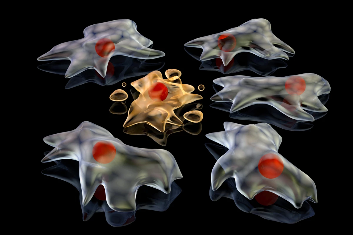
When cells undergo apoptosis, a programmed form of death, they emit signals that attract immune cells, known as phagocytes, to clean up cellular debris. While scientists have identified several signals, including “find-me” and “eat-me” signals, the new study introduces a physical trace that adheres to surfaces and directs immune cells to the precise location of cell death. The researchers have designated this trace as FOOD, short for Footprint of Death.
Professor Ivan Poon, head of the laboratory at the La Trobe Institute for Molecular Science, emphasized the significance of this finding: “Understanding this basic biological process could open new avenues of research to develop new treatments that harness these steps and help the immune system better fight disease.” With billions of cells dying daily as part of normal body functions, the understanding of this process is crucial for advancing medical research.
Utilizing advanced techniques such as microscopy, live-cell imaging, and proteomics, the team studied how FOOD is formed and its behavior. Researchers induced both human and mouse cells to undergo apoptosis and observed the retraction of cell bodies, leaving behind a distinct footprint rich in F-actin and other adhesion proteins. Unlike most extracellular vesicles that detach, FOOD remains anchored to the surface where the cell died.
The study found that FOOD appeared in nearly all tested cell types and under various triggers for cell death. Once a cell leaves its footprint, it begins to form vesicles, termed FOOD-derived extracellular vesicles (F-ApoEVs). These vesicles signal immune cells with the classic “eat-me” cue, prompting them to engulf the remnants.
The process relies on the activity of ROCK1, a protein that governs cell contraction. When ROCK1 function was inhibited, FOOD formation was disrupted. In total, the proteomic analysis identified 601 proteins within FOOD, primarily those associated with cell structure and adhesion, indicating that FOOD comprises mainly membrane and cytoskeletal components.
In experiments, immune cells known as macrophages effectively recognized and cleared F-ApoEVs when placed near FOOD. This interaction appears to “prime” macrophages for enhanced clearance of other dying cells. Notably, in cells infected with influenza A, FOOD and F-ApoEVs were found to contain viral proteins and complete viral particles, suggesting that these vesicles can act as reservoirs for viral spread.
This dual functionality indicates that while FOOD assists the immune system in locating dead cells, it can also be exploited by viruses to propagate infections. Lead author Stephanie Rutter, a PhD candidate in Poon’s lab, noted, “What we didn’t expect was how viruses can also take advantage of this process and cause infection by hiding in F-ApoEVs.”
Co-author Georgia Atkin-Smith, a senior postdoctoral researcher at the Walter and Eliza Hall Institute of Medical Research, added, “This study has revealed that dying cells can continue to communicate from the grave and may impact immune function.”
Despite the valuable insights, the study does have limitations. Most experiments were conducted in vitro, leaving the behavior of FOOD in living organisms uncertain. Additionally, the formation of FOOD was primarily observed in adherent cells, raising questions about its role in free-floating cells, such as those in blood. The long-term fate of FOOD structures in tissues remains unclear, and findings related to viral spread are specific to influenza A, indicating that other viruses may behave differently.
Understanding the mechanisms behind FOOD could offer new strategies for promoting tissue repair and managing inflammation. Further research into the role of FOOD may lead to innovative therapies that enhance immune responses and improve health outcomes. As Rutter concluded, “The more we understand about cell death and what happens to cells after they die, the better we can understand disease pathologies and find new treatments.”
The full findings are documented in the journal Nature Communications.

Schumer and Advocates Protest 70% Cuts to Housing Program

Bull Shark Attack Claims Life and Leaves Man Seriously Injured

Navigating Friendship: How to Address Allergies with Tact

Wharton Professor Reveals 5 Mindset Shifts for Working Moms

Astronics Receives Upgrade to Strong-Buy Amid Positive Earnings

Experts Warn of Rising Crisis as Global Female Prison Population Nears One Million

Skoda Reimagines Classic Sedan with Futuristic Electric Design
Oklahoma Lawmakers Urged to Enhance Charter School Regulations

How Sarah Josepha Hale Paved the Way for Thanksgiving

Discover the Top 10 Calorie Counting Apps of 2025

Bella Hadid Shares Health Update After Treatment for Lyme Disease

Erin Bates Shares Recovery Update Following Sepsis Complications

Discover 2025’s Top GPUs for Exceptional 4K Gaming Performance

Electric Moto Influencer Surronster Arrested in Tijuana

Discover How to Reverse Image Search Using ChatGPT Effortlessly

Meta Initiates $60B AI Data Center Expansion, Starting in Ohio

Recovering a Suspended TikTok Account: A Step-by-Step Guide

Tested: Rab Firewall Mountain Jacket Survives Harsh Conditions
-
 Technology4 months ago
Discover the Top 10 Calorie Counting Apps of 2025
-
 Health2 months ago
Bella Hadid Shares Health Update After Treatment for Lyme Disease
-
 Health3 months ago
Erin Bates Shares Recovery Update Following Sepsis Complications
-
 Technology4 weeks ago
Discover 2025’s Top GPUs for Exceptional 4K Gaming Performance
-
 Technology2 months ago
Electric Moto Influencer Surronster Arrested in Tijuana
-
 Technology4 months ago
Discover How to Reverse Image Search Using ChatGPT Effortlessly
-
 Technology4 months ago
Meta Initiates $60B AI Data Center Expansion, Starting in Ohio
-
 Technology4 months ago
Recovering a Suspended TikTok Account: A Step-by-Step Guide
-
 Health4 months ago
Tested: Rab Firewall Mountain Jacket Survives Harsh Conditions
-

 Lifestyle4 months ago
Belton Family Reunites After Daughter Survives Hill Country Floods
-

 Technology3 months ago
Uncovering the Top Five Most Challenging Motorcycles to Ride
-

 Technology4 months ago
Harmonic Launches AI Chatbot App to Transform Mathematical Reasoning